●
Orthopedics: Restore bone translocation, reset, fixing.
●
Surgery: Taking foreign bodies out of the body, cardiac catheterization, implantable pacemakers, interventional treatment, angiography, and local photography.
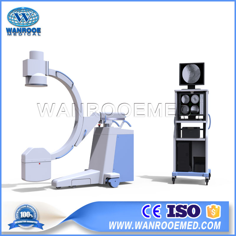
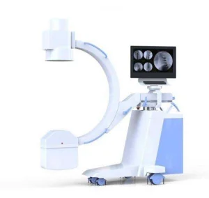
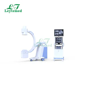
C-arm mainframe (1 set)
X-ray generator & Power supply (5.0kW, 40 kHz)
Imported 9 inch image intensifier
Mega-pixel digital radiography system
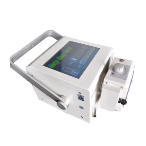
Digital acquisition workstation
Imported dense grain grids
Adjustable beam limiting device
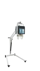
19 inch medical LCD grayscale displays (2 sets)
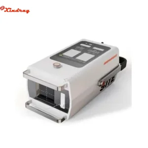
Hand-held controller (1 set)
Frequently Asked Questions (FAQ)
Q1: What are the primary medical applications for the PLX112B?
The PLX112B is primarily used in orthopedics for bone fixing and resetting, as well as in general surgery for foreign body removal, cardiac catheterization, and interventional treatments.
Q2: How does the pulse fluoroscopy feature benefit surgery?
Pulse fluoroscopy provides high-precision, clearer images at a much lower radiation dose, which significantly enhances safety for both the medical staff and the patient during complex minimally invasive procedures.
Q3: Is the equipment compatible with hospital data systems?
Yes, it features a standard DICOM interface, making it highly convenient to exchange information and images with existing hospital management systems.
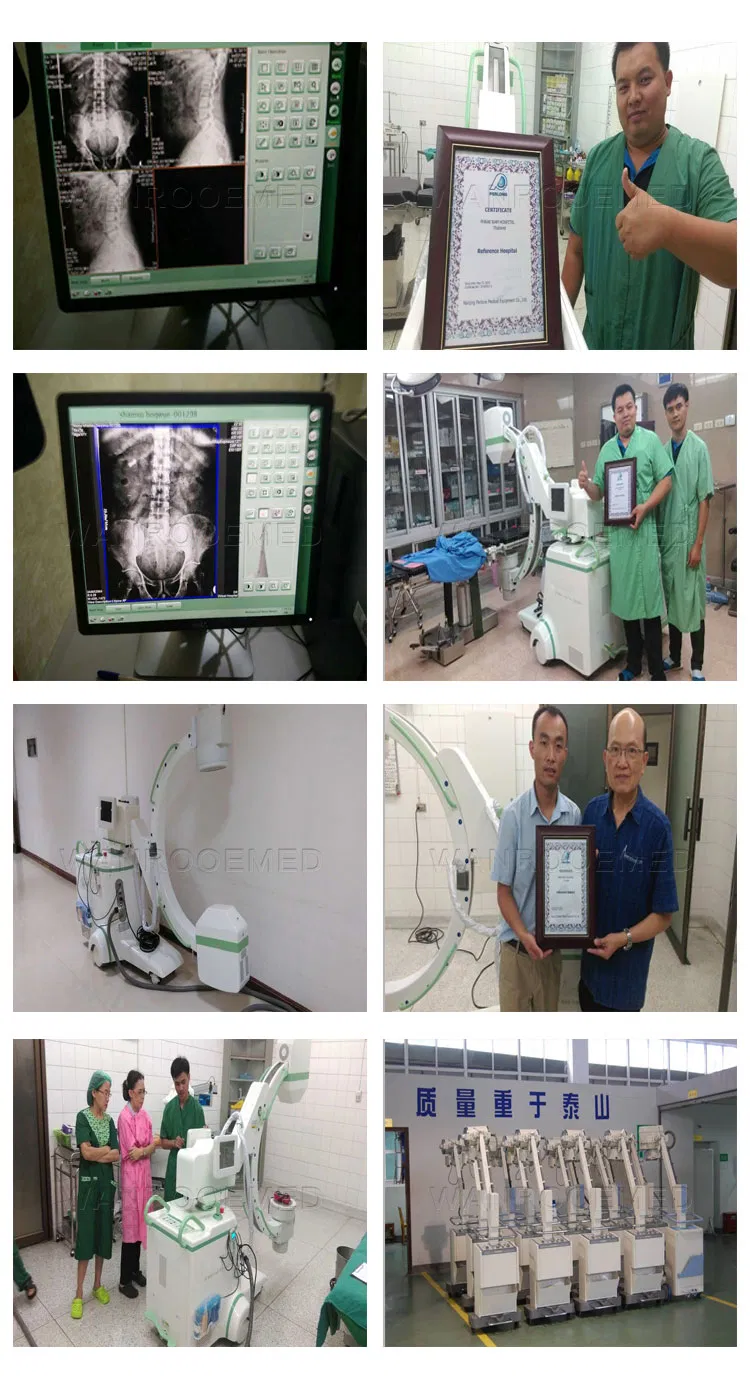
Q4: What is the benefit of the dual-monitor design?
The dual monitors allow for synchronized observation inside and outside the operating room, facilitating better teamwork and real-time surgical teaching or consultation.
Q5: What imaging technology is used in the PLX112B?
It utilizes an imported 9-inch image intensifier combined with a mega-pixel ultra-low illumination digital CCD photography system to ensure high-stability and high-clarity images.
Q6: Can the C-arm be easily adjusted during operation?
Yes, it features a unique hand-held controller and an electric auxiliary support arm design, allowing for smooth movement and precise control over radiation fields and parameters.
