📋 Basic Information Overview
| Model NO. | THR-XR-8500C |
| Use as | High Frequency Digital Radiography System |
| Power Output | 55 Kw |
| Rotary Anode Speed | 8400rpm |
| mAs Range | 0.1~630mas |
| Inverter Frequency | 60 kHz |
| Tube Voltage | 40~150kv |
| AEC Control | Shortest time 5ms |
| Thermal Capacity | 900kj (1200 Khu) |
| Tube Current | 10-500mA |
| Power Supply | 380V 50Hz |
| Specification | CE and ISO |
| Origin | China |
| HS Code | 9022140090 |
| Production Capacity | 50000PCS/Year |
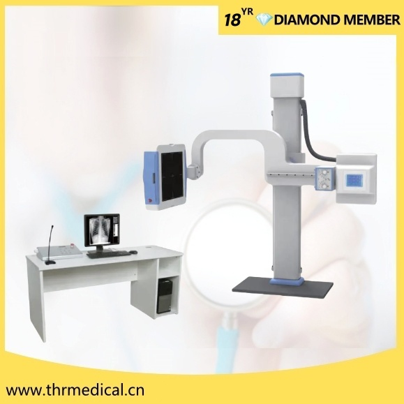
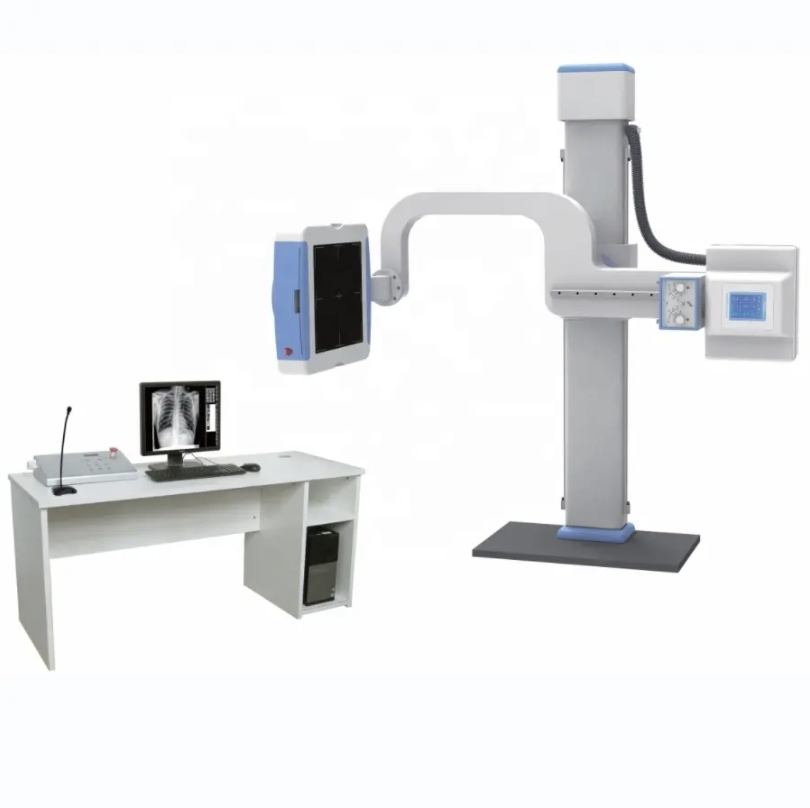
🔍 Product Description
THR-XR-8500C
High Frequency Digital Radiography System (55KW, 500mA)
💻 Image Workstations Capabilities
-
⚙️
Acquisition Module: Gigabit network acquisition with adaptive image enhancement processing.
-
🖼️
Image Processing: Includes photo processing, X-ray synchro control, and motion control software. Features tissue equilibrium, W/L adjustment, Gamma correction, noise reduction, sharpening, and edge extraction.
-
💾
Storage & Transmission: Supports DICOM direct transmission, DICOM Worksite SCU, standard DICOM, DIR, and film printing.
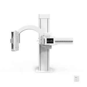
🌟 Key Features
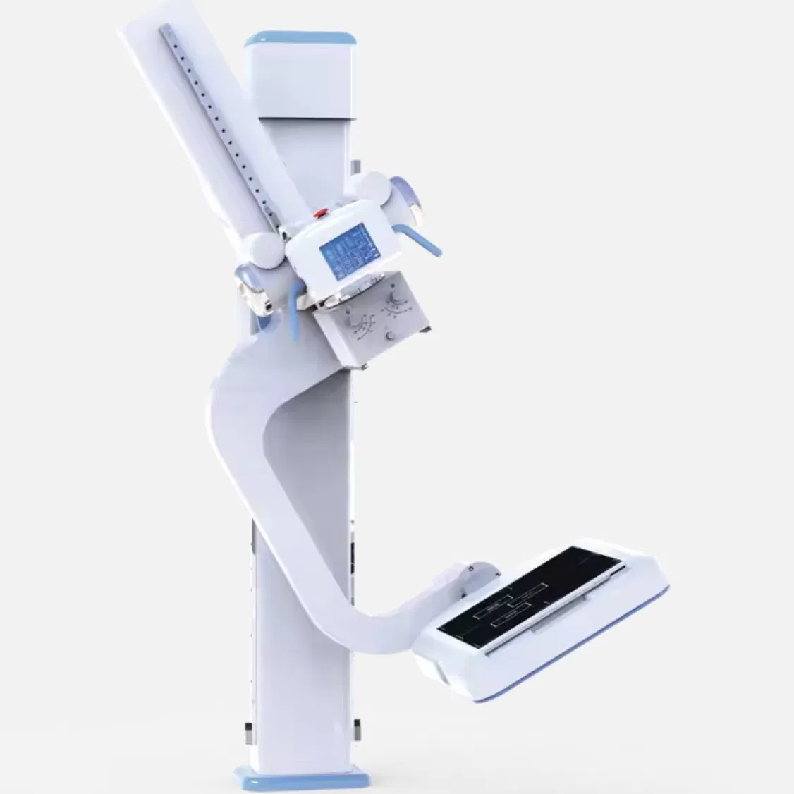
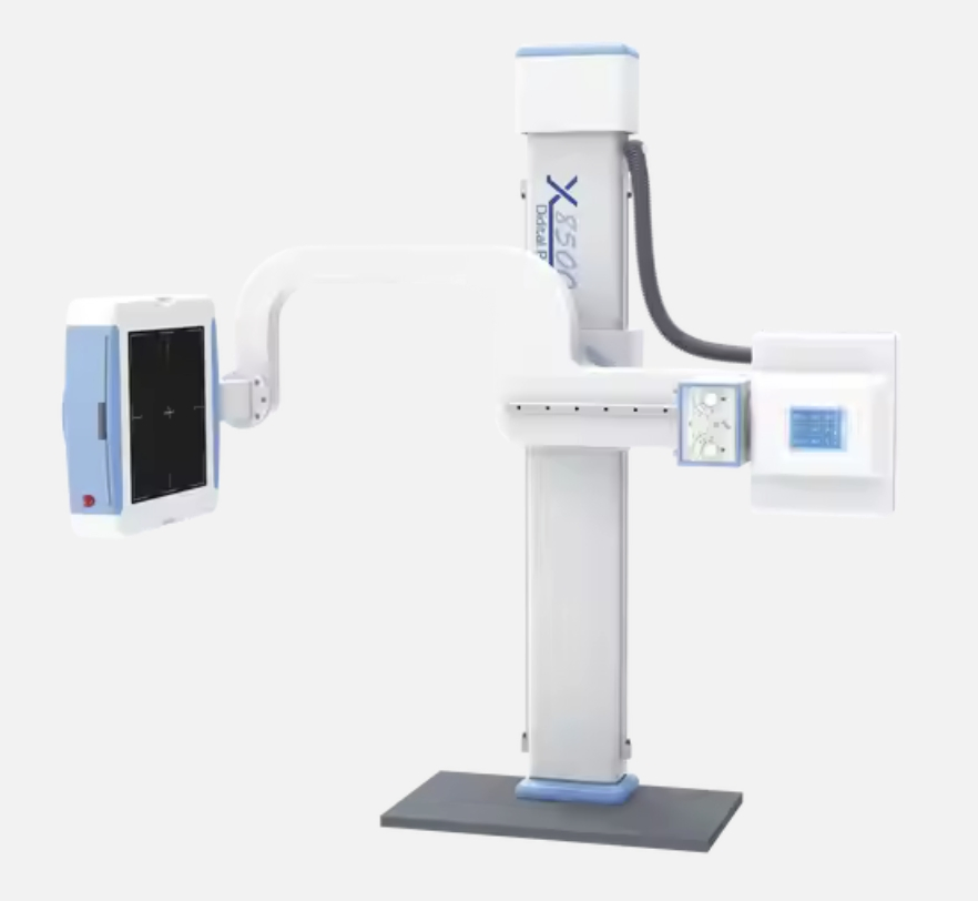
- 🔹Amorphous silicon flat detector with advanced manufacturing for super stability.
- 🔹Large 17" x 17" effective detection area to meet all photographic needs.
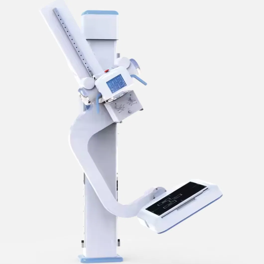
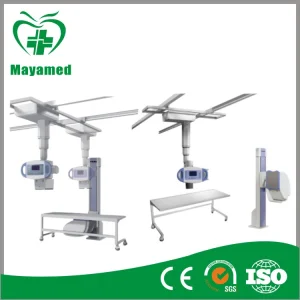
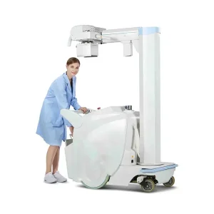
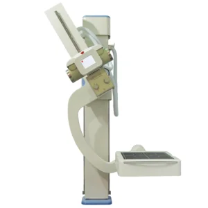
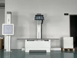
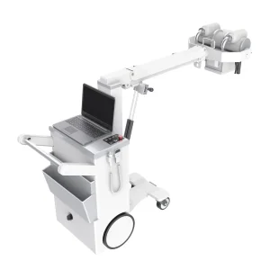
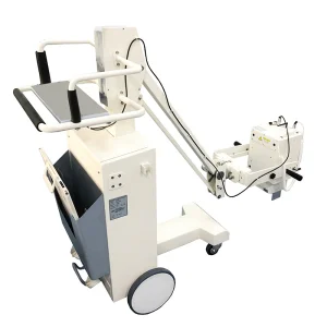
- 🔹Electric lift and rotatable U-shaped frame for flexible standing and lying positions.
- 🔹Compact high-frequency X-ray generator eliminates the need for extra high-voltage cables.
- 🔹Floating photographic bed with electromagnetic locks for accurate patient positioning.
- 🔹Intelligent touchable LCD control system with graphic real-color interface.
- 🔹Digital closed-loop control of KV and MA for precise dose repeatability.
- 🔹Multiple automatic protection features and fault tips for operational safety.
📊 Technical Parameters
Electrical Properties
| Power Output | 55 KW |
| Inverter Frequency | 60 kHz |
| Dual-focus | Small: 0.6 / Large: 1.2 |
| Thermal Capacity | 900kJ (1200 KHU) |
| Rotary Anode Speed | 8400rpm |
| Tube Voltage | 40~150kV |
| Tube Current | 10-500mA |
| mAs | 0.1~630mAs |
| AEC | Shortest time 5ms |
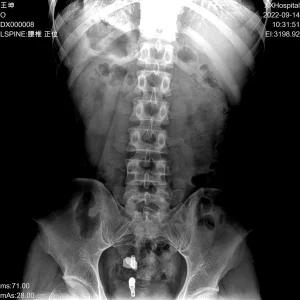
Digital Image Processing System
| Detector Type | Amorphous silicon flat detector |
| Scintillator Type | Cesium Iodide (CsI) |
| Field of View | 17 * 17 Inch |
| Pixel Matrix | 3k × 3k |
| Spatial Resolution | 3.6Lp/mm |
| Pixel Size | 143μm |
| Grayscale Output | ≥14bits |
| DQE | ≥70% |
| Imaging Time | ≤12s |
🏢 Manufacturing Excellence
❓ Frequently Asked Questions
What are the primary advantages of the U-shaped frame design?
The U-shaped frame is designed for maximum flexibility, featuring electric lift and rotation. This allows medical professionals to easily perform radiography for patients in various positions, including standing, sitting, or lying down, making the workflow more efficient.
Does the system support integration with hospital PACS networks?
Yes, the system fully supports DICOM 3.0 standards. This ensures seamless connectivity with PACS (Picture Archiving and Communication Systems) for easy image transmission, storage, and remote printing.
How does the system ensure patient safety regarding radiation dose?
The system utilizes high-frequency inverter technology and digital closed-loop control. Combined with the AEC (Automatic Exposure Control) which can react in as little as 5ms, it ensures high-definition images are produced with the lowest possible radiation dose.
What type of detector technology is used in this DR system?
The THR-XR-8500C uses an advanced Amorphous Silicon flat panel detector with a Cesium Iodide (CsI) scintillator. This combination provides high DQE (Detective Quantum Efficiency) and excellent spatial resolution for superior image quality.
What are the power requirements for installing this equipment?
The system requires a standard industrial power supply of 380V at 50Hz. The high-frequency generator is integrated into the design to save space and simplify installation.
How long does it take to see the image after exposure?
The digital imaging system is highly efficient, with a total imaging time of 12 seconds or less, allowing for rapid diagnosis and high patient throughput.


