Pipmed Medical Pipmed Medical

10 Tips to Optimize Imaging Systems for Hospitals

Time:2026-10-29 Author:Sophia
0%

Hospital imaging departments rarely struggle because of one scanner alone. Delays often begin earlier: an incomplete order, a poorly timed appointment, or a room that sits idle while staff search for supplies. The World Health Organization has reported more than 3.6 billion diagnostic radiological examinations worldwide each year, underscoring the scale of the service hospitals must coordinate. Yet volume alone does not reveal local bottlenecks. Leaders need to examine their own scheduling, equipment utilization, image quality, staffing, and patient handoffs before choosing solutions.

A useful systems principle comes from physician and healthcare quality-improvement expert Paul Batalden: “Every system is perfectly designed to get the results it gets.” Applied to imaging, that idea shifts attention from individual effort to workflows, data, and everyday constraints. How long does a patient wait between registration and scan? How often does a scanner remain unused because of preventable downtime? Small details matter. A missing protocol can delay a whole list. So can an unrealistic appointment template. This guide explores how to optimize imaging systems for hospitals through practical steps, measurable indicators, and careful investment decisions. It also recognizes an uncomfortable truth: optimization can expose old assumptions, and not every proposed improvement will work on the first try. Hospitals should test changes, monitor safety and access, and revise plans using real operational data—not just vendor promises.
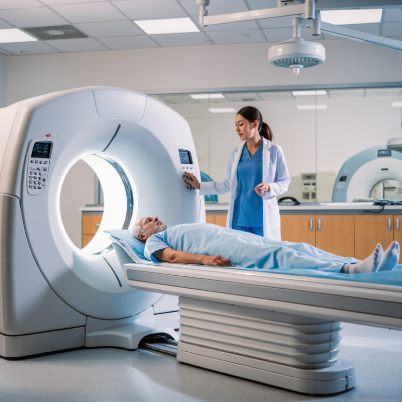
10 Tips to Optimize Imaging Systems for Hospitals

Assess Imaging Needs, Capacity, and System Performance

A hospital should begin with its actual imaging workload, not a wish list of new equipment. Map referrals by modality, time of day, urgency, and patient type. Then compare demand with staffed scanner hours, maintenance downtime, and room availability. OECD’s Health at a Glance 2023 reported an average of about 17 MRI units per million people across OECD countries, with wide variation between countries. That gap matters. A national average cannot tell a hospital whether its evening MRI queue is caused by too few slots, staffing gaps, or avoidable repeat scans.

Measure performance at each step: order-to-scan time, cancellations, room turnover, report turnaround, and repeat-image rates. Break results down by modality and shift; a hospital-wide average can hide a Monday morning bottleneck. UNSCEAR’s 2020/2021 report estimated roughly 4.2 billion diagnostic radiological examinations worldwide in 2016. CT made up about one-tenth of procedures while contributing a much larger share of collective radiation dose. That makes protocol review and dose tracking part of capacity planning, not a separate task. The uncomfortable caveat: utilization figures can look efficient while staff are rushing or patients wait in corridors. Review a few real patient journeys with technologists and radiologists; spreadsheets miss details. Reassess after schedule changes, equipment upgrades, and seasonal demand shifts. Not just volume.

Improve Equipment Placement and Patient Workflow

Imaging efficiency often begins with the walk from the entrance to the scanner. Place registration and changing areas near the appropriate imaging rooms, while keeping waiting patients away from active transport routes. A wheelchair should turn without blocking a doorway. Staff should be able to move a stretcher from the corridor to the room with minimal repositioning. Small details matter.

Map the full patient journey, including check-in, preparation, scanning, and discharge. Observe it during busy periods, not only when rooms are quiet. If patients repeatedly ask where to go, signs or sightlines may need attention. Keep clean supplies close to the point of use, but preserve clear access to equipment and emergency exits. Storage can quietly become a bottleneck.

A floor plan can look efficient on paper and still feel confusing in practice. Ask technologists, nurses, transport staff, and patients where delays occur; their perspectives may differ. Test proposed changes with temporary signs or movable furniture before committing to construction. Not every path can be shortened. Privacy, staff communication, and safe transfers may matter more than saving a few steps. Review the layout after changes, because workflows shift as services and patient needs change.

Integrate Imaging Systems with Hospital IT

Integrating imaging with hospital IT means more than connecting scanners to a picture archive. A chest X-ray should arrive in the correct patient record, with its order and clinical notes attached. Connect modality worklists, radiology information systems, picture archiving systems, and electronic health records using established DICOM and HL7 interfaces. Where available, use FHIR APIs for appropriate data exchange. Test patient matching carefully; a technically successful transfer can still put an image in the wrong chart. Small errors matter.

The 2023 ONC report on interoperability found that 70% of U.S. hospitals engaged in all four measured exchange activities: sending, receiving, finding, and integrating electronic information. That figure is not specific to imaging, but it shows why integration must include usable data, not just transmission.

Build a test environment with sample orders, canceled exams, and corrected patient details. Monitor interface queues and record failures, then assign staff to resolve them. Keep downtime procedures visible near reading rooms.

One uncomfortable truth: interfaces need ongoing maintenance, and hospitals may underestimate the staff time this takes. Test again after software updates; assumptions age quickly.

Enhance Image Quality, Safety, and Preventive Maintenance

Image quality, patient safety, and reliable maintenance depend on measurements, not guesswork. NCRP Report No. 160 estimated that U.S. medical radiation exposure averaged about 3.0 mSv per person in 2006, up from 0.53 mSv in 1980. This historical figure is not a current hospital benchmark, but it shows why dose deserves regular review. Compare local CT dose data with diagnostic reference levels. ICRP Publication 135 describes setting these levels using the 75th percentile of dose surveys. A level is a prompt to investigate, not a target to reach by lowering exposure blindly. Small changes matter.

Build practical checks into routine work. Review image artifacts, repeat rates, exposure settings, and quality-control results by scanner and protocol. A faint ring on a phantom image may signal detector drift; a sudden rise in repeats may point to positioning or workflow problems. Record the finding, action, and follow-up date. Preventive maintenance should include scheduled inspections, calibration checks, and timely service when performance falls outside established tolerances. Keep the evidence. Equipment logs can reveal gradual deterioration that a single inspection misses. Still, checklists are imperfect: busy teams may document a test without acting on a borderline result. Assign a named owner to review trends, and verify that corrections improve both image quality and dose.

Monitor Results and Plan Future Upgrades

A hospital’s imaging system should be judged by more than its age or appearance. Track repeat-scan rates, room downtime, report turnaround, and patient wait times each month. A rising repeat rate may point to unclear instructions, inconsistent positioning, or a protocol that needs review. Check image quality with radiologists and technologists, not numbers alone. Context matters.

Keep a simple dashboard that staff can actually use. For example, compare a Monday morning’s equipment delays with the service log and staffing notes. Record what changed, who reviewed it, and whether the problem returned. Small samples can mislead, and busy teams may enter data late. That happens. Flag gaps rather than treating incomplete records as firm evidence.

Use these findings to shape upgrade plans before equipment becomes unreliable. List recurring faults, parts availability, software support needs, and compatibility with existing systems. Ask users which steps create delays, such as transferring studies or preparing patients. Then rank upgrades by clinical impact, operational risk, and cost. Leave room for uncertainty: one month of poor performance may reflect a temporary staffing issue, not a failing system. Revisit priorities with clinical, technical, and financial teams as new data arrives.

FAQS

How can imaging departments improve patient flow?

Place check-in and changing areas near the relevant rooms. Keep waiting patients clear of stretcher routes. A wheelchair needs room to turn.

Why observe patient journeys during busy periods?

Busy periods reveal delays that quiet rooms hide. Watch check-in, preparation, scanning, and discharge. Repeated questions may signal confusing signs or sightlines.

How should teams test layout changes?

Try temporary signs or movable furniture before construction. Ask technologists, nurses, transport staff, and patients where delays happen. Their answers may differ.

What should imaging system integration include?

Link scanners with patient records, exam orders, and clinical notes. Test sample orders, cancellations, and corrected patient details. Check every patient match.

How can hospitals catch integration failures?

Monitor interface queues and assign staff to resolve errors. Keep downtime instructions near reading rooms. Interfaces need maintenance. Assumptions age quickly.

Which measures help assess imaging performance?

Track repeat scans, room downtime, report turnaround, and patient waits each month. Review image quality with clinical staff, not numbers alone.

How should teams interpret performance data?

Compare equipment delays with service logs and staffing notes. Flag missing records instead of treating them as firm evidence. Small samples can mislead.

What should guide future equipment upgrades?

Consider recurring faults, parts availability, software support, compatibility, clinical impact, operational risk, and cost. One poor month may reflect staffing, not failing equipment. Revisit priorities as new data arrives.

Conclusion

Hospitals can improve imaging services by first assessing clinical needs, patient volumes, equipment capacity, and system performance. This helps identify bottlenecks and determine whether existing resources are being used effectively. Thoughtful equipment placement, clear scheduling, and coordinated patient workflows can reduce delays and make imaging visits more efficient for both patients and staff.

A practical approach to how to optimize imaging systems for hospitals also includes connecting imaging tools with hospital information systems, protecting reliable access to images, and supporting smooth communication among care teams. Consistent image-quality checks, radiation-safety practices, and preventive maintenance help keep equipment dependable and patient-focused. By monitoring service outcomes, equipment use, and maintenance needs, hospitals can spot opportunities for improvement and plan future upgrades based on changing clinical demands.

Sophia

Sophia

Sophia is a dedicated marketing professional with an exceptional depth of knowledge about her company's products and services. With a keen understanding of market trends and customer needs, she crafts insightful blog posts that not only inform but also engage readers, enriching the company’s online......