Pipmed Medical
In 2026, the question “what is the role of imaging in early detection” deserves a careful, evidence-based answer. Imaging can reveal subtle changes before symptoms become obvious. A low-dose CT scan may show a tiny lung nodule. Mammography can identify architectural distortion. MRI can expose tissue changes that ultrasound may miss. These findings can create valuable time for confirmation, treatment, and informed decisions.
Yet imaging is not a crystal ball. Its value depends on the right test, suitable timing, technical quality, and skilled interpretation. Radiologists also consider age, symptoms, family history, previous scans, and possible risks. A bright spot on an image may represent disease, inflammation, or harmless variation. That uncertainty matters. False positives can lead to anxiety, extra procedures, and unnecessary costs. False negatives can delay care.
Curtis Langlotz, MD, PhD, a Stanford radiologist and medical imaging researcher, has said, “AI will not replace radiologists, but radiologists who use AI will replace radiologists who don’t.” His statement highlights a practical direction for modern imaging. Artificial intelligence may help prioritize urgent scans, compare images over time, and detect patterns that human eyes sometimes overlook. However, it should support—not replace—clinical judgment, patient discussion, and established diagnostic pathways. The strongest approach combines advanced equipment with experienced professionals and transparent evidence. Small technical errors can matter. So can overconfidence. Early detection works best when imaging is used thoughtfully, verified clinically, and explained clearly to the person behind the scan.
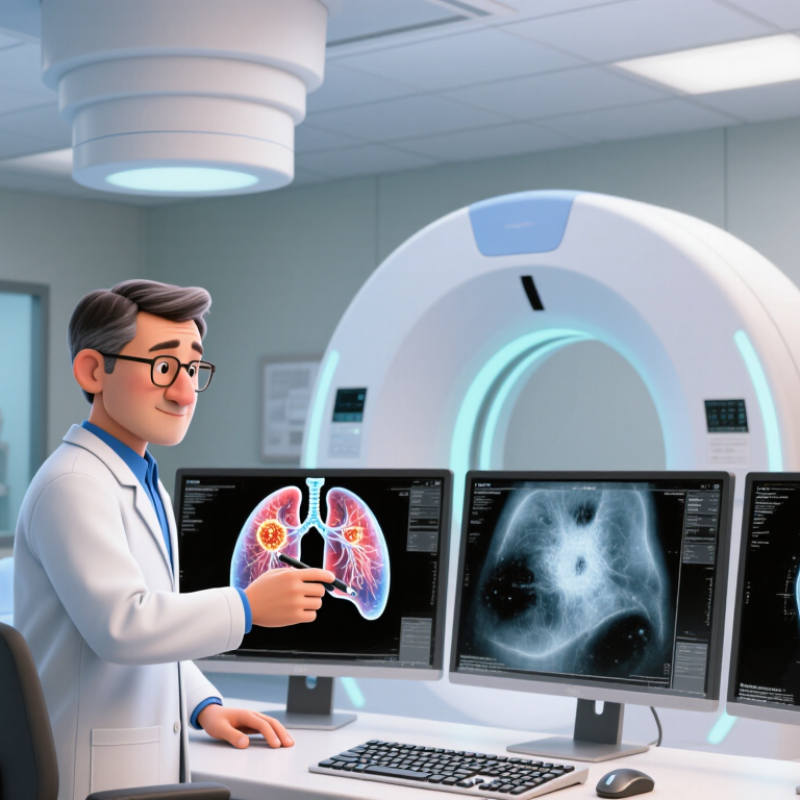
Early Detection Imaging Means in Modern Healthcare
Early detection imaging uses medical scans to identify disease before symptoms become obvious. In 2026, clinicians may use low-dose CT, MRI, ultrasound, or mammography, depending on a person’s risk and symptoms. Each method reveals different details. A scan can show a tiny lung nodule, a blocked artery, or an unusual tissue pattern. These findings can guide earlier treatment and reduce uncertainty during difficult clinical decisions.
However, early detection is not automatically better. It is not flawless. Some findings are harmless but trigger repeat scans, biopsies, or anxiety. Radiation exposure also matters, especially when scans are unnecessary or repeated too often. Experienced clinicians should consider age, family history, previous results, and the patient’s concerns before recommending imaging. Evidence-based guidelines can support that decision, but they cannot replace careful conversation.
A patient may remember the waiting room more clearly than the scan itself. Clear explanations can improve trust. The report should describe what was found, what remains uncertain, and what action is reasonable. Artificial intelligence may help detect subtle changes, yet trained professionals must review its suggestions. Access is another concern: advanced imaging can be expensive or unavailable outside major hospitals. Modern healthcare should pursue earlier answers without treating every shadow as disease.
Imaging supports early detection by identifying disease before symptoms appear. The chart shows the recommended starting ages for selected population-based imaging screening programs in average-risk adults under current United States preventive-care guidance.
These recommendations apply to specific populations and risk profiles. Individual screening decisions should consider personal history, risk factors, benefits, and potential harms.
Sources: U.S. Preventive Services Task Force recommendations for breast cancer, lung cancer, colorectal cancer, and abdominal aortic aneurysm screening.
Early detection often begins before symptoms become noticeable. Modern imaging helps clinicians see structural, functional, and molecular changes inside the body. Low-dose computed tomography can reveal tiny lung nodules during carefully selected screening. Magnetic resonance imaging can show detailed tissue contrast without ionizing radiation. Ultrasound displays blood flow and soft tissue movement in real time. These tools do not diagnose disease alone. Radiologists compare images with medical history, physical examinations, and laboratory findings.
Some techniques detect altered metabolism before an organ changes shape. This can guide further testing when suspicious areas appear. Image quality depends on motion control, timing, equipment, and reader experience. A clear scan is not always a clean bill of health. Small lesions may remain invisible, while harmless findings can create anxiety. That uncertainty matters. Doctors must weigh benefits, radiation exposure, contrast reactions, and follow-up needs.
In practice, the best examination matches the person and the clinical question. A family history may justify closer evaluation, but it does not prove disease. Shared decisions improve trust when results remain unclear. Artificial intelligence may highlight patterns, yet human review remains essential. Early imaging works best as one step in a broader care pathway. It should prompt informed action, not automatic treatment. Research continues to refine screening thresholds and reduce unnecessary procedures.
Imaging helps clinicians find disease before symptoms become severe. The best method depends on the body area, risk level, and clinical question.
Plain X-rays remain useful for fractures, chest infections, and some lung changes. They are quick, but small or early abnormalities may remain hidden.
Ultrasound uses sound waves and avoids ionizing radiation. It often guides evaluation of gallbladder disease, thyroid nodules, pregnancy, and soft-tissue masses. It can also assess blood flow when Doppler imaging is added.
Computed tomography creates detailed cross-sectional images. It is valuable for suspected stroke, internal bleeding, lung nodules, and complex injuries. However, radiation exposure matters, especially during repeated examinations.
Magnetic resonance imaging provides excellent detail for the brain, spine, joints, and pelvic organs. It can reveal subtle tissue changes that X-rays may miss.
Mammography supports breast cancer screening, while low-dose chest CT may help selected people with a high smoking-related risk. Positron emission imaging can show active disease, but it usually works best when combined with other evidence.
No scan is perfect. A clear image does not always exclude illness, and an unexpected finding may cause anxiety or unnecessary testing. This part is easy to underestimate. Clinicians should match imaging to symptoms, medical history, examination findings, and established guidelines. Patients also deserve a plain explanation of benefits, limitations, radiation exposure, and possible follow-up before the scan.
2026 Top Imaging for Early Detection: What Is the Role?
Choosing an imaging test is less like picking the newest tool and more like asking a precise clinical question. A doctor considers symptoms, timing, medical history, age, and examination findings. Chest pain may require a rapid study, while a slowly changing lump may need another pathway. Context matters most. Early detection depends on matching the test to the suspected problem, not scanning everything.
Ultrasound can show movement and soft tissues without radiation, but body size and gas may reduce its clarity. Computed tomography can provide detailed images quickly, especially during emergencies. However, it uses radiation and may require contrast material. Magnetic resonance imaging offers excellent soft-tissue detail, yet it can take longer and feel difficult for some patients. Each method has limits.
Before ordering a scan, clinicians review previous images to avoid unnecessary repetition. They check kidney function, allergies, implanted devices, pregnancy status, and the urgency of symptoms. A radiologist’s interpretation also matters; the image is not the diagnosis by itself. No imaging choice is perfect. Even experienced doctors may revise the plan when symptoms change or an image remains unclear. Patients can ask what question the test should answer, what risks exist, and what happens after the result. Sometimes the safest choice is a different test, a short follow-up, or careful observation.
Early detection imaging can reveal disease before symptoms become obvious. In clinical practice, that timing may open more treatment options. Low-dose computed tomography can identify small lung nodules, while magnetic resonance imaging can show soft-tissue changes without ionizing radiation. Ultrasound may offer a practical option during pregnancy or in regions with limited resources. Each method answers different questions, so the “top” test depends on age, symptoms, family history, and risk level.
Real clinics teach caution. Imaging can produce false positives, incidental findings, and unnecessary procedures. Radiation exposure matters, especially after repeated examinations. Contrast agents can also cause reactions or create concerns for patients with kidney problems. A scan does not equal a diagnosis. Qualified clinicians must interpret results alongside medical history and physical findings. Clear communication is essential, because alarming images can create anxiety before evidence is complete. It is tempting to call every new scan revolutionary. That confidence can be misplaced.
Future developments may combine faster scanners, lower radiation doses, and artificial intelligence that highlights subtle patterns. These tools could support earlier review, but they should not replace professional judgment. Reliable systems need testing across different ages, ethnic groups, body types, and healthcare settings. Privacy protection must remain central when medical images train algorithms. Portable imaging may expand access, yet availability alone will not guarantee accurate care. Human oversight, transparent reporting, and shared decisions should guide adoption. Some uncertainty will remain. That is not failure; it is a reason to monitor outcomes carefully.
Imaging can reveal disease before symptoms become severe. It supports earlier clinical decisions. A clear scan can still miss illness.
Plain X-rays are often used for fractures, chest infections, and some lung changes. They are quick and widely available. Small abnormalities may remain hidden.
Ultrasound can assess the gallbladder, thyroid, pregnancy, blood flow, and soft-tissue lumps. It uses sound waves, not ionizing radiation. Body gas or larger body size may reduce image clarity.
Computed tomography can quickly examine suspected stroke, internal bleeding, lung nodules, and complex injuries. It provides detailed cross-sectional images. Radiation exposure still matters.
Magnetic resonance imaging provides strong detail for the brain, spine, joints, and pelvic organs. It may show subtle tissue changes. The scan can take time.
Mammography supports breast cancer screening and may identify changes before symptoms appear. Screening decisions should follow personal risk and clinical guidance.
Selected people with high smoking-related risk may be considered for low-dose chest computed tomography. It is not suitable for everyone.
No. A clear image does not always exclude illness. Symptoms, examination findings, and medical history still matter. This limitation is easy to underestimate.
Doctors may review previous images, kidney function, allergies, implanted devices, pregnancy status, and symptom urgency. Repeated scans are not automatically better.
Ask what clinical question the test should answer, what risks exist, and what happens afterward. Sometimes observation is safer. The best choice is not always obvious.
Early detection imaging plays an important role in modern healthcare by helping clinicians identify disease before noticeable symptoms develop. The question “what is the role of imaging in early detection” can be answered by examining how different technologies reveal structural, functional, or molecular changes inside the body. Imaging methods may support the early evaluation of conditions affecting the brain, heart, lungs, breasts, bones, and other organs, with each technique offering specific strengths and limitations.
Doctors choose an imaging test according to the patient’s symptoms, medical history, age, risk factors, and the information needed for diagnosis. Early imaging can support faster treatment decisions, improve monitoring, and sometimes reduce the need for more invasive procedures. However, potential concerns such as radiation exposure, contrast reactions, false-positive findings, cost, and access must be considered. Future developments are expected to improve image quality, personalized screening, automated analysis, and the ability to detect subtle changes earlier while maintaining patient safety.